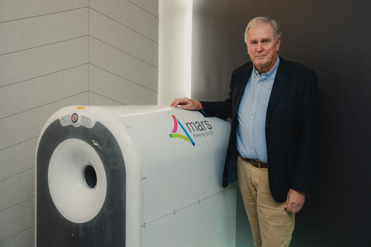
Above: Professor Phil Butler with the MARS 5x120 Extremity spectral CT in a clinic
When Professor Phil Butler began his BSc(Hons) at the University of Canterbury back in 1965, little did he realise that he would become one of its longest serving academic staff members.
The highly acclaimed scientist retired in late 2021 after a stellar career spanning 50 plus years. During his tenure, he supervised more than 40 PhD students, made an extraordinary commitment to research, inspired a love of Science & Technology in school children – and was even instrumental in setting up UC’s first creche for use by staff.
Phil held various roles within UC, including Head of Department in 1997-1998 and 2002-2006. He served as Pro-Vice-Chancellor (Resources) 1998-2000 and as Pro-Vice-Chancellor (Services) 2000-2001.
Phil’s dedication to research was never self-serving. Most important to him was the desire to contribute something to the global knowledge base while at the same time passing his depth of knowledge onto his students and inspiring a new generation of scientists.
“For me, research has always been about contributing to positive changes to society. I see little point in research unless it is used.
“Seeing a small number of physicists and chemists use my algebraic methods and tables used for calculations in crystal structure, in LED technology, in nuclear models and in particle physics is motivating. But seeing hundreds of thousands of New Zealand school kids enjoying Science & Technology classes and exhibitions is very motivating.”
Phil was a driving force behind the establishment of the National Science-Technology Roadshow Trust that has toured Science & Technology (S&T) exhibitions to mostly primary and intermediate schools around New Zealand since 1988. It continues to attract upwards of 50,000 visitors annually.
In 1991 the Trust opened Science Alive, the “S&T museum” in the old Christchurch Railway Station. It was destroyed by the 2010/2011 earthquakes. Science Alive currently provides free online S&T lessons tailored to the Kiwi school syllabus.
Phil was one of four academics who, also in 1991, persuaded the then Vice-Chancellor to establish Canterprise, which was charged with commercialising university inventions.
“It really changed some views within UC that for research to be taken up by industry, UC needed to be active and take the initiative. This acceptance by UC that scientists should strive to have societal impact from their research has been gratifying,” Phil says.
Case in point is the world’s first 3D colour x-ray CT machine which Phil invented with son Anthony, previously at UC and now a professor at Otago University. It is being commercialised by UC’s spinout company, MARS Bioimaging Ltd. The revolutionary scanners will enable doctors to make more accurate diagnoses when treating everything from broken bones to heart disease.
There are 300 million CT scans performed each year around the world as part of medical care. MARS and a small number of other groups have established that spectral photon counting will be the routine way to do CT for the next few decades.
“It’s particularly gratifying that the knowledge gained from this MARS research will have such a broad impact,” Phil says.
“This means I have played a small part in helping a very large number of people. More locally, it's been gratifying to develop a technology that has already earned millions of dollars in export revenue, and over the next few years this is likely to grow significantly.”
Clinicians have been able to use MARS for decision-making since last year. MARS is now accredited by relevant New Zealand health funding agencies. Clinical trials are under way in New York.
Phil says it’s been particularly gratifying to work with his son on MARS research and in the company MARS Bioimaging Ltd.
“It was an extremely broad set of aims and I believe our very close working relationship, coupled with different science perspectives, enabled us to jointly make progress across such diverse topics – with progress often faster than other research groups.”
Professor Anthony Butler shares his father’s satisfaction.
“Few people are lucky enough to work closely with a family member. I've been extremely lucky to work with my father on cutting edge science. Having jointly run a project spanning physics, engineering, and medicine to develop a medical imaging tool is a rare opportunity for a son or daughter. I'm really proud that our combined efforts are already improving the medical care for our community.
“Often it’s been the little things Phil did that are an important benefit for UC staff. When I was working at UC, I often encountered staff who were sending their young children to the staff creche on Ilam road. I've proudly told them that my father was involved in setting up the creche. It is really nice knowing that my father's impact to UC was more than just research and teaching – rather it also involved supporting other staff in a broad number of ways.”
Although now retired, Phil is still fully occupied. He works part-time at MARS Bioimaging, remains a trustee of Science Alive, and continues to think about algebraic issues in Mathematical Physics.
“I’m still too busy, life is still varied, fun, full and challenging.”

.jpeg)
